
Anatomy becomes difficult when learners must convert flat illustrations into an accurate mental picture of structures that occupy three-dimensional space. A nerve may pass behind a vessel, a muscle may cover several deeper tissues, and an organ may change appearance when viewed from another plane. Modern anatomy teaching therefore depends not only on naming structures but also on showing how they relate, overlap, and connect. A virtual dissection system gives educators another way to make those relationships visible while preserving the systematic organization required in medical education.
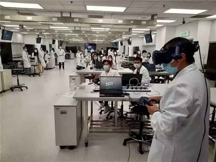
From Static Images to Spatial Anatomy
Textbooks and projected slides remain useful for introducing terminology, but each image presents anatomy from a predetermined viewpoint. Learners must mentally combine multiple illustrations before they can understand depth or orientation. A virtual dissection table presents high-resolution anatomical models that can be enlarged, rotated, and observed from different directions.
These operations are particularly useful when a structure changes course through the body. Students can follow a vessel across regions, examine the path of a nerve, or compare superficial and deep structures without losing the surrounding context. Instead of memorizing isolated pictures, they can study anatomy as a connected spatial system.
The platform does not remove the need for anatomical terminology. Rather, it connects names with positions, shapes, and neighboring structures, giving each term a clearer physical meaning.
Interaction as a Learning Method
Touch-based control changes the learner’s role from passive observer to active investigator. On a digital dissection platform, bones, muscles, nerves, vessels, and other structures can be manipulated directly. A class can begin with the intact body, remove selected layers, rotate the model, and then restore the original view for another demonstration.
Repeated interaction supports comparison. A learner can inspect a structure from an anterior view, switch to a lateral perspective, and then examine a sectional image of the same area. If the spatial relationship remains unclear, the sequence can be repeated without physically damaging a specimen..
The educational value comes from purposeful manipulation rather than movement alone. Each rotation, cut, or layer removal can be tied to a specific question, such as which tissue lies deeper or which route a vessel follows.
Connecting Different Branches of Anatomy
Medical programs commonly divide anatomy into systemic, regional, and sectional study. A virtual dissection table can connect these approaches within the same digital environment. Systemic anatomy may trace the nervous or vascular system through the whole body, while regional anatomy concentrates on all structures within areas such as the thorax or pelvis. Sectional anatomy then examines how those structures appear in transverse, sagittal, or coronal planes.
Moving between these views is valuable because clinical imaging rarely presents structures exactly as they appear in a textbook diagram. When students compare a three-dimensional model with sectional information, they begin to recognize why an organ or vessel has a particular shape in a CT or MRI image.
This integrated method also gives instructors flexibility. DIGIHUMAN’s digital anatomy resources can support an introductory lecture, a focused regional lesson, or an imaging-based case discussion..
Repetition Without Physical Degradation
Cadaveric dissection provides direct experience with real human tissues and natural variation, but specimens are limited and require specialized procurement, preservation, storage, and disposal. A digital anatomy platform provides reusable anatomical models that can be restored to their original state after each session..
Repeated practice is especially useful during revision. Different groups can perform the same sequence of observations, and an instructor can return to an earlier stage without reconstructing a physical dissection. DIGIHUMAN’s reusable digital format also avoids direct exposure to formaldehyde and the biohazard concerns associated with preserved material.
These advantages do not mean that every physical teaching method becomes unnecessary. The two formats address different educational needs. Cadaveric work provides tactile and hands-on experience with real human tissues, while digital exploration offers repeatability, controlled visualization, and rapid movement between anatomical views..
A Structured Place in the Modern Curriculum
Technology contributes most when it follows a clear teaching objective. Instructor-led demonstrations, small-group exploration, pre-laboratory preparation, and post-laboratory review can all be organized around a virtual dissection table. Educators can identify structures, hide selected anatomical layers or structures, create sectional views, and ask learners to explain the relationships revealed by each operation..
The platform can also support discussion rather than individual screen use alone. A group may predict what lies beneath a visible layer before it is removed or compare two possible routes for a nerve. Assessment can follow the same principle by asking students to locate a structure, describe its neighbors, or interpret its appearance in another plane rather than simply recall a label.
Because the model can be returned to a prepared state, instructors can present comparable exercises to several groups. Consistent starting conditions make discussion more focused while still leaving room for learners to explain different observational routes.
DIGIHUMAN places this teaching approach within a software-and-hardware platform built around interactive digital human data. Its resources include real anatomical videos and animations, test-question materials, tomographic data, and corresponding CT and MRI images. Modern anatomy education still requires accurate content, expert instruction, and carefully chosen activities. By making anatomical layers and spatial relationships repeatable, visible, and open to guided investigation, the technology gives educators a practical connection between traditional anatomical knowledge and contemporary medical learning.